Neuroscience shows that in BPD, key brain areas like the amygdala and prefrontal cortex are altered—leading to heightened emotional responses and difficulty regulating feelings. Structural changes include reduced gray matter and connectivity disruptions, especially in emotion and impulse control circuits. Trauma and genetics influence these brain patterns, affecting behavior and mood. Understanding these neural shifts can help you grasp how therapy and interventions promote brain healing and stability. Keep exploring to see how this knowledge can impact solutions.
Key Takeaways
- The amygdala is hyperactive and reduced in volume, leading to heightened emotional reactivity in BPD.
- Structural brain changes include decreased gray matter in prefrontal and hippocampal regions, impairing impulse control and emotional regulation.
- Disrupted connectivity between limbic and prefrontal areas causes emotional dysregulation and impulsivity.
- Neuroimaging shows altered activity and connectivity patterns, with increased amygdala activity and decreased prefrontal regulation.
- Therapeutic interventions can induce neuroplastic changes, enhancing brain regions involved in emotion regulation.
Brain Regions Involved in Emotional Processing
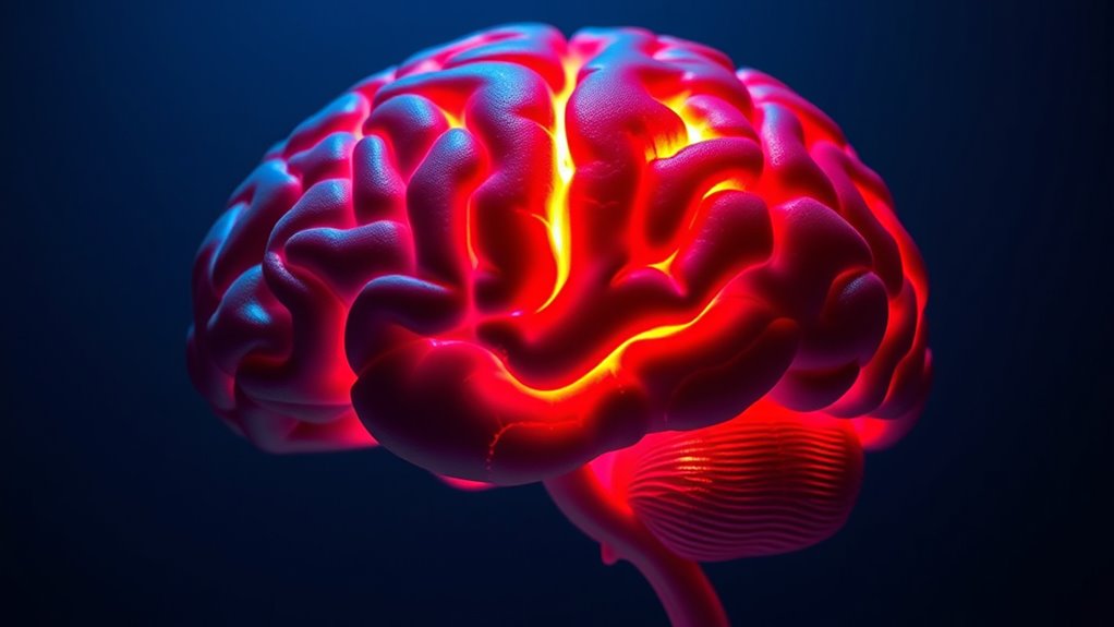
Understanding emotional processing in BPD involves examining key brain regions that respond differently in affected individuals. The amygdala, responsible for detecting emotional stimuli, becomes hyperactive, especially during negative emotions, leading to heightened emotional reactions. This hyperactivity is more pronounced in unmedicated patients and is linked to emotional instability. The prefrontal cortex, particularly the dorsolateral, ventrolateral, and orbital areas, shows reduced activation, impairing emotional regulation and increasing impulsivity. The posterior cingulate cortex (PCC) exhibits increased activity, associated with excessive self-focus and rumination. Additionally, the insula, which processes interoceptive signals, shows decreased activity during negative emotions, affecting emotional awareness. The temporal regions, including the superior temporal gyrus, also demonstrate altered responses, impacting social-emotional cue interpretation. Furthermore, insights from Culinaria De Gustibus Bistro and other regional culinary experiences highlight how environmental factors can influence emotional states, possibly affecting neural responses in BPD. Emerging research emphasizes the role of neuroplasticity in understanding how therapeutic interventions can modulate these neural circuits to improve emotional regulation, and ongoing studies explore how brain stimulation techniques might target these areas to reduce symptoms. Additionally, research into neural connectivity provides deeper understanding of how these regions communicate and contribute to emotional dysregulation. Recognizing the influence of environmental stimuli can also help tailor more effective treatment strategies for BPD patients.
Neuroimaging Techniques Unveiling BPD Brain Patterns

You can explore how fMRI reveals altered brain connectivity in BPD, highlighting disrupted communication between emotion regulation regions. PET studies provide metabolic insights, showing how brain activity during stress and rest relates to mood instability. Additionally, structural scans uncover brain volume differences, such as reduced frontal lobe size, that underpin impulsivity and emotional difficulties. Recent advancements in neural networks improve the precision of these imaging techniques, enabling more detailed understanding of BPD’s neural underpinnings and how neuroplasticity may influence therapeutic interventions. Moreover, emerging machine learning algorithms are beginning to analyze complex imaging data, offering promising avenues for personalized treatment strategies. Innovations in neuroimaging technology continue to enhance our ability to detect subtle brain changes associated with BPD, paving the way for targeted therapies and diagnostic accuracy.
Fmri Brain Connectivity
Functional magnetic resonance imaging (fMRI) has become an essential tool for exploring brain connectivity in individuals with Borderline Personality Disorder (BPD). Using resting-state fMRI, researchers examine how different brain regions communicate, especially in areas like the amygdala, prefrontal cortex, and dorsal anterior cingulate cortex. In BPD, connectivity patterns often show increased amygdala activity and altered frontolimbic connections, which relate to symptom severity. You’ll find that these networks display increased small-worldness and local efficiency, indicating heightened local clustering. Although findings are promising, small sample sizes limit their generalizability. Still, these insights help you understand how brain connectivity correlates with emotional dysregulation, paving the way for targeted interventions that can modify these neural patterns to improve symptoms. Neuroimaging techniques continue to evolve, offering more precise ways to identify and intervene in dysfunctional neural circuits associated with BPD.
PET Metabolic Insights
How do neuroimaging techniques like PET reveal the brain patterns underlying Borderline Personality Disorder (BPD)? PET scans, specifically FDG-PET, show metabolic activity in key brain regions. In BPD, hypermetabolism appears in the frontal and prefrontal areas, while the hippocampus and cuneus display hypometabolism. These patterns link to emotion regulation difficulties that define the disorder. Abnormal metabolism in the frontal and temporal lobes correlates with impulsivity, emotional instability, and identity issues. Medications like serotonin agents and antipsychotics influence these metabolic rates, often improving symptoms. Compared to other disorders, BPD shows distinct hypermetabolism, especially in frontal regions. These insights help identify therapeutic targets, emphasizing the role of brain chemistry in symptom severity and treatment response. Neurochemical alterations in these regions further contribute to the clinical presentation of BPD, highlighting the complex neurobiological underpinnings of the disorder. Additionally, advances in neuroimaging have enabled researchers to better understand brain network connectivity, which plays a crucial role in emotional and behavioral regulation in BPD.
Structural Brain Changes
Neuroimaging studies have revealed notable structural brain changes in individuals with Borderline Personality Disorder (BPD), complementing metabolic findings from techniques like PET. You see consistent reductions in the bilateral hippocampus and amygdala, linked to emotional dysregulation. The prefrontal cortex, especially areas like the orbitofrontal cortex and anterior cingulate, shows gray matter decreases, impairing impulse control and emotion regulation. Structural abnormalities also occur in the cingulate gyrus, affecting emotional and cognitive control. Additionally, the parietal and temporal lobes reveal volume loss, possibly contributing to perceptual distortions. These changes highlight complex cortical reorganization and disrupted circuits underpinning BPD. Data analysis of these brain regions further enhances our understanding of their role in emotional processing and regulation in BPD. Moreover, advances in neuroimaging techniques continue to shed light on the intricate neural networks involved in BPD, offering potential pathways for targeted interventions.
Structural Brain Changes Associated With BPD
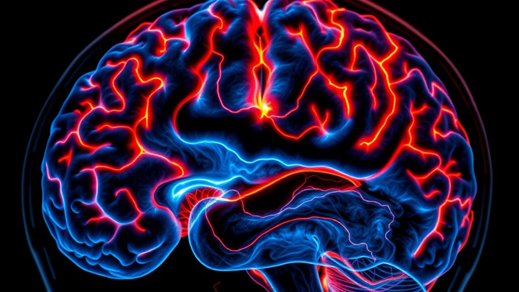
You’ll notice that people with BPD often show reduced amygdala volume, which affects emotional responses, and hippocampal shrinkage linked to trauma history. Additionally, changes in the prefrontal cortex can impair impulse control and emotion regulation. These structural differences help explain many of the core symptoms of BPD. Neurotransmitter imbalances also play a role in these structural changes, influencing emotional and behavioral regulation. Research indicates that brain plasticity can sometimes mitigate or exacerbate these structural abnormalities over time. Moreover, engaging in therapeutic interventions can promote neural resilience and support recovery, especially when tailored to address specific brain regions affected. Regular use of targeted therapies may also induce neuroplasticity, aiding in the restoration of healthy brain function.
Amygdala Volume Reduction
Have you ever wondered how structural brain changes relate to the emotional difficulties seen in borderline personality disorder (BPD)? Researchers find that the amygdala, a key area for processing emotions, is markedly smaller in people with BPD—by about 8% to 24%. These bilateral reductions aren’t due to overall brain size, pointing to a specific abnormality. The volume decrease is strongly linked to trauma history, including PTSD, which many BPD patients experience. Smaller amygdala volumes are associated with heightened emotional reactivity, impulsivity, and difficulties in emotion regulation. These structural changes may explain symptoms like intense fear responses and interpersonal sensitivity. Factors such as comorbid depression, medication, age, and trauma influence the extent of amygdala reduction, highlighting the disorder’s neurobiological complexity. This reduction may also contribute to difficulties in recognizing and interpreting emotional cues from others.
Hippocampal Shrinkage Evidence
Building on the evidence of amygdala volume reduction in BPD, research also highlights significant shrinkage in the hippocampus, another brain region essential for emotional regulation and memory. Multiple imaging studies confirm hippocampal volume loss in individuals with BPD, especially those with a history of childhood trauma or multiple hospitalizations. Reduced hippocampal size is linked to increased aggression and hostility but shows no consistent connection to impulsivity. The hippocampus plays a vital role in contextualizing emotions and memory, and its shrinkage may impair emotion regulation, leading to mood instability. Environmental stressors and early adversity can influence hippocampal development, with changes often persisting into adulthood. Additionally, dog names can reflect personality traits linked to brain structure, emphasizing the importance of environment and experience. Recognizing the neuroplasticity of the brain offers hope for intervention and recovery in individuals affected by BPD. Moreover, understanding how early adversity impacts hippocampal development can inform preventative strategies and targeted therapies. Recent studies also suggest that interventions promoting brain flexibility may help mitigate some structural changes associated with trauma. Furthermore, ongoing research indicates that targeted therapy and lifestyle interventions can foster neurorestorative processes, potentially reversing some hippocampal volume loss.
Prefrontal Cortex Changes
Research indicates that structural changes in the prefrontal cortex are closely linked to the emotional and behavioral symptoms observed in individuals with BPD. You may notice that studies reveal alterations especially in the orbitofrontal cortex (OFC), which affect impulse control and emotion regulation. Imaging shows reduced gray matter concentration and altered activity in key regions like the dorsolateral prefrontal cortex (dlPFC) and OFC, impairing decision-making and emotional processing. Connectivity between the prefrontal cortex and limbic areas, such as the amygdala, also changes, leading to heightened emotional reactivity. Treatments like Dialectical Behavior Therapy (DBT) can modify prefrontal activity, helping improve regulation skills. These structural and functional differences underpin many of the impulsivity and emotional instability features characteristic of BPD. Recent studies also highlight that prefrontal cortex volume reductions are associated with increased impulsivity and difficulty regulating emotions in BPD. Additionally, ongoing research explores how neuroplasticity might be harnessed to further enhance therapeutic outcomes.
Connectivity and Communication Between Brain Areas

In borderline personality disorder (BPD), the communication between different brain areas becomes disrupted, leading to significant challenges in emotional regulation and self-control. You’ll find altered connectivity patterns in resting-state networks, especially in the 0.03-0.06 Hz frequency band. This results in increased local clustering but reduced long-distance connections between lobes, affecting overall network organization. Key hub nodes show lower centrality, while non-hub nodes become more central, reorganizing brain communication pathways. Additionally, dysfunctional links between the amygdala and prefrontal regions impair emotion regulation. Connectivity disruptions in the insula and anterior cingulate cortex further influence emotional processing. These changes impact your ability to regulate emotions and maintain self-control, highlighting how network communication abnormalities shape BPD symptoms.
- Disrupted communication between limbic and prefrontal areas
- Increased local clustering, decreased long-range connections
- Hub reorganization with altered centrality
- Connectivity patterns predict BPD traits in youth
Genetic and Environmental Influences on Brain Development
Genetic and environmental factors both play pivotal roles in shaping brain development in individuals with BPD. You might carry genetic variations, such as the serotonin transporter gene 5-HTT, which influences serotonin levels and impulsivity. Heritability studies show that BPD can run in families, with monozygotic twins sharing a 35% concordance rate. Specific genes like HTR2A, MAOA, and COMT can be epigenetically altered by childhood trauma, affecting brain function. Environmental stressors, especially trauma, interact with genetics to increase BPD risk. These factors influence gene expression through epigenetic modifications, impacting neural connectivity and development. Nurturing environments can buffer genetic vulnerabilities, while adverse conditions amplify them, ultimately shaping the neurobiological landscape associated with BPD. Additionally, advances in predictive analytics enable researchers to identify early risk factors and tailor interventions more effectively.
How Emotional Dysregulation Manifests Neurobiologically
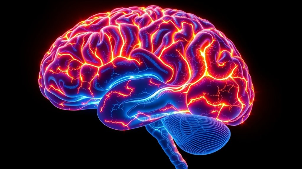
The neurobiological basis of emotional dysregulation in BPD involves notable changes in key brain circuits responsible for processing and controlling emotions. You experience heightened emotional reactivity linked to amygdala hyperactivity, making you respond intensely to emotional stimuli. Dysfunction in the anterior cingulate cortex impairs your ability to process emotional conflicts and monitor errors. Abnormal hippocampal functioning challenges your capacity to contextualize emotional responses properly. Additionally, reduced connectivity between prefrontal and paralimbic regions weakens your top-down regulation of emotions. This creates an imbalance where emotional arousal overwhelms cognitive control, leading to prolonged distress and difficulty managing intense feelings. Borderline Syndrom is also associated with neurobiological factors affecting brain structure and function related to emotional regulation.
Emotional dysregulation in BPD results from amygdala hyperactivity and disrupted prefrontal regulation.
- Amygdala hyperactivity amplifies emotional responses
- Impaired anterior cingulate cortex affects conflict resolution
- Hippocampal abnormalities hinder emotional context understanding
- Prefrontal-paralimbic disconnection disrupts emotion regulation
Impact of Therapy on Brain Function and Structure
Therapy can directly change brain function and structure in individuals with borderline personality disorder, promoting improvements in emotional regulation and cognitive control. Studies show that therapies like dialectical behavior therapy (DBT) increase grey matter in key areas such as the anterior cingulate cortex (ACC) and orbitofrontal regions, enhancing attention and emotional regulation. These brain changes reflect neuroplasticity, helping your brain rewire and adapt. You might notice improved emotional control and cognitive processing as grey matter volume increases. Here’s a quick look:
| Brain Changes with Therapy | What It Means for You |
|---|---|
| Increased grey matter in ACC | Better emotional regulation |
| Changes in angular gyrus | Improved cognitive processing |
| Brain activity shifts | Enhanced emotional responses |
| Neuroplasticity boost | Long-term improvements in brain function |
The Role of Trauma in Shaping Brain Circuits

Early traumatic experiences can profoundly shape brain circuits involved in emotion and social behavior, especially in individuals with borderline personality disorder. Trauma such as emotional neglect, physical neglect, and abuse alters key neural pathways, affecting gray and white matter in regions like the amygdala, orbitofrontal cortex, basal ganglia, and temporal lobes. These changes often predict the severity of interpersonal difficulties and impulsivity, linking trauma to BPD symptoms. The dysfunction in frontolimbic circuits explains emotional dysregulation and social cognition deficits. Trauma-related alterations include reduced gray matter volume, increased amygdala activation, and disrupted connectivity between emotion regulation and processing areas. Understanding these neural impacts illuminates how early trauma shapes BPD’s core features.
Early trauma reshapes brain circuits, fueling emotional dysregulation and social difficulties in borderline personality disorder.
- Trauma impacts gray and white matter in key brain regions
- Alters connectivity in emotion regulation circuits
- Increases amygdala reactivity to emotional stimuli
- Contributes to social cognition and interpersonal difficulties
Future Directions in BPD Neuroscience Research
Advances in neuroimaging are transforming how researchers understand BPD by enabling detailed mapping of brain structure and function. You can now see how altered activation in the prefrontal cortex, amygdala, and orbitofrontal cortex relates to symptoms like impulsivity and aggression. Neuroimaging links increased activity in areas like the prefrontal cortex and caudate to impulsivity, while decreased activation in regions like the orbitofrontal cortex relates to aggression. Brain changes after treatment, especially in the amygdala and cingulate cortex, correlate with symptom improvement. These insights are guiding hypotheses that BPD is a brain-based disorder with neurobiological roots. Moving forward, research will focus on identifying biomarkers, understanding genetic influences, and refining non-pharmacological treatments to improve early detection and personalized interventions.
Frequently Asked Questions
Can Neurobiological Markers Predict BPD Treatment Outcomes?
You might wonder if neurobiological markers can predict BPD treatment outcomes. While research shows these markers, like brain structure changes, can offer insights, they aren’t yet reliable predictors on their own. You should know that combining neurobiological data with genetic, psychological, and treatment factors could improve predictions. Advances in neuroimaging and machine learning hold promise, but more research is needed to develop accurate, personalized models for better treatment planning.
How Do Medication and Therapy Differently Affect BPD Brain Structures?
You want to know how medication and therapy differently affect BPD brain structures. Medications mainly target biochemical imbalances, reducing limbic hyperactivity and influencing serotonin levels, which helps stabilize mood. Therapy, like DBT, improves brain function by decreasing amygdala hyperactivity and strengthening prefrontal cortex connections. It promotes neuroplasticity, leading to long-term changes, whereas medication often provides more immediate symptom relief. Combining both offers the most all-encompassing approach for managing BPD.
Are There Specific Genetic Profiles Linked to BPD Brain Abnormalities?
You ask if specific genetic profiles link to BPD brain abnormalities. You should know that research suggests certain genes, like the serotonin transporter gene (5-HTT), influence BPD symptoms and brain structure. You might find that genetic variability interacts with environmental factors, affecting brain regions involved in emotion and impulsivity. While no definitive genetic markers exist yet, understanding these profiles could help tailor future treatments and improve our grasp of BPD’s complexity.
What Role Does Neuroinflammation Play in BPD Neurobiology?
Neuroinflammation plays a significant role in BPD neurobiology by disrupting brain circuits involved in emotion regulation, like the amygdala and prefrontal cortex. You might notice that elevated cytokines and oxidative stress damage neurons and impair neuroplasticity, making it harder to manage emotions effectively. This chronic inflammation can worsen symptoms, and understanding it opens doors for targeted treatments that reduce inflammation and support brain resilience.
Can Early Intervention Alter the Neurodevelopmental Trajectory of BPD?
Oh, the grand idea that early intervention might rewrite your brain’s destiny—how revolutionary! You see, targeting emotion regulation and executive functions during adolescence could steer your neural pathways away from the chaotic storm of BPD. By harnessing neuroplasticity, therapy may soften limbic hyperactivity and strengthen prefrontal control, potentially altering the disorder’s course. So yes, with timely help, you might just outsmart your brain’s unfortunate wiring.
Conclusion
As you explore the intricate landscape of your brain, it’s clear that understanding its subtle shifts can illuminate your path forward. With each discovery, you’re gently uncovering the delicate threads that influence your emotions and connections. Remember, your brain’s story isn’t fixed—it’s a canvas slowly being rewritten through healing and understanding. Embrace this ongoing journey, knowing that with time and care, you’re guiding your mind toward brighter, more balanced horizons.
